About me / Research

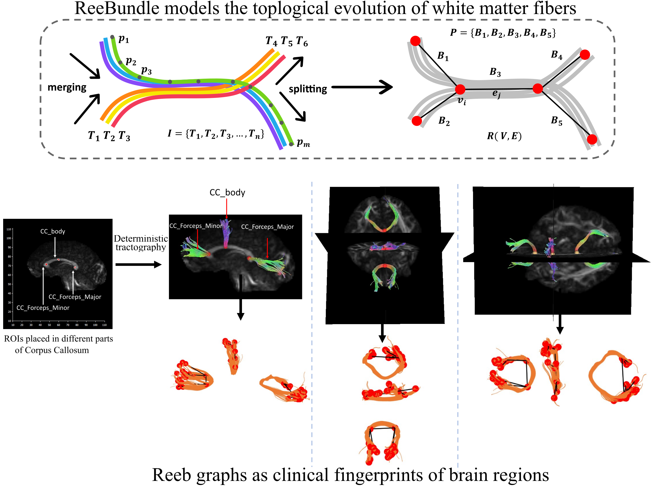
Brain Connectivity Analysis

Our brain is a complicated anatomical network responsible for cognition and behavior. Neurodegenerative diseases impede the associated network architecture in millions of affected people. My research aims to mathematically model the neuronal activities in the human brain using tools from computational geometry and artificial intelligence to characterize the evolution of neuronal fibers. We model the high-level topological structures of the fibers from diffusion MRI imaging data to construct graph-based mathematical objects. My research will pave the way for advanced AI methods to compare brain regions and provide pathological insights that are currently infeasible due to the complexity of the neuronal fibers.

Analysis of the shunt treatment for NPH

The diagnosis of Normal Pressure Hydrocephalus (NPH) through quantitative analysis of ventricular volume has been largely under-explored and the changes in ventricular volume post-shunt surgery have not been fully quantified. Radiologists rely on visual examination of CT scans which may not accurately capture the changes in the volume. This study shows the effectiveness of a ventricular volume metric, computed using deep learning models, in differentiating NPH patients from normal individuals with high accuracy.

Segmentation / Classification

Segmenting different regions in glioma is a challenging task. Current state-of-art method uses UNet and DeepMedic architecture and attains the maximum accuracy of about 88%. In this paper, we propose a method that adds an average ensembling layer on top of the existing neural network model which has the best known performance. We show that this improves the accuracy of tumor segmentation to 90%. Also, we show results from different methods by varying the ensembling layer and its classifiers that suggest an improvement in the accuracy. We show that leveraging the uniqueness of each model is an important step towards building predictive models. We use the BraTS2018 dataset for all our experiments.
We present a method of nuclei boundary refinement in our algorithm that takes advantage of this boundary adherence feature in SLIC. Later, to track the segmented nuclei, we propose a graph-based tracking method for linking cell nuclei frame-by-frame. Our cell segmentation algorithm is based on a few key observations of N3DH-CE: (1) considerable variation in size and staining intensity of the embryonic nuclei, (2) the lack of sufficient gold truth for segmentation, and (3) availability of weakly annotated data for detection.